The first X-ray image of an animal, specifically a frog, was made by Arthur Schuster, a British physicist of German origin. At that time, however, this was still only a physics experiment. The use of imaging diagnostic methods in veterinary medicine began only decades later, between the two World Wars. The first professional book on the subject, Veterinary Radiology by Paul Henkel, was published in Berlin in 1926, with 226 pages and 91 illustrations. In 1927, with American support, an X-ray Institute was established at the Vienna University of Veterinary Medicine, which at that time still operated as a college.
In those early years, only a small number of veterinarians had the opportunity to take X-ray images. The widespread use of the method began after the Second World War, partly due to technological progress and partly because of the growing focus on the treatment of small animals.
This method has been supporting the work of our Zoo’s veterinarians for more than half a century, and today it would be hard to imagine daily practice without it. While in the past we used X-ray film that had to be developed, modern X-ray machines now work with digital sensors. This allows images to be viewed immediately on a monitor and saved right away.
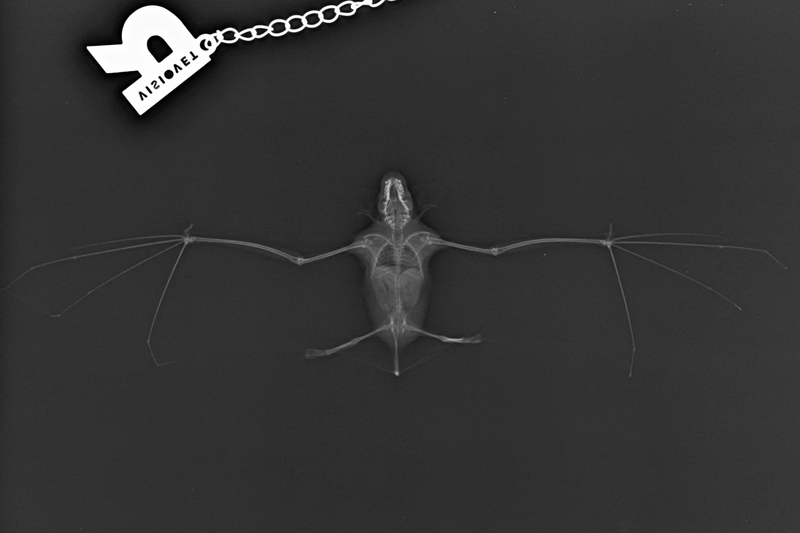
X-rays are useful primarily for diagnostics, as they allow veterinarians to look inside the body without major intervention and determine exactly what the problem is. This is important not only in the treatment of our own animals, but also in the care of the 2,000 to 3,000 rescued animals that arrive each year at the Zoo’s Wildlife Rescue Centre. Even images taken of healthy animals can be interesting, as they clearly show anatomical features, especially the skeletal structure. This is particularly relevant in our Zoo, where many species have anatomical characteristics that differ in several ways from the body structure of more familiar animals such as dogs, cats, or other commonly kept pets.